HRV Introduction and the importance of regulation
The heart as a success organ, is controlled in its function by the autonomic nervous system and continuously adapted to the current situation!
“The constant adjustment of the intervals from heartbeat to heartbeat (RR spikes) by the superior autonomic nervous system enables the organism to react in fractions of a second to changed life situations in order to survive = constant regulation”
The biologically meaningful reactions to life-threatening events (formerly fight-or-flight) are today transferred and interpreted to many everyday situations such as: Partnership stress, quarrels with your own children, job stress, trouble with colleagues or the boss, the constant feeling of being overwhelmed in everyday situations, letters from the “tax office”, frequent watching of exciting TV shows, movies or sports or playing action video games etc..
Sympathetic and parasympathetic nervous systems ultimately control all organs, tissues and cells. Result: normal function, “overfunction” or “underfunction
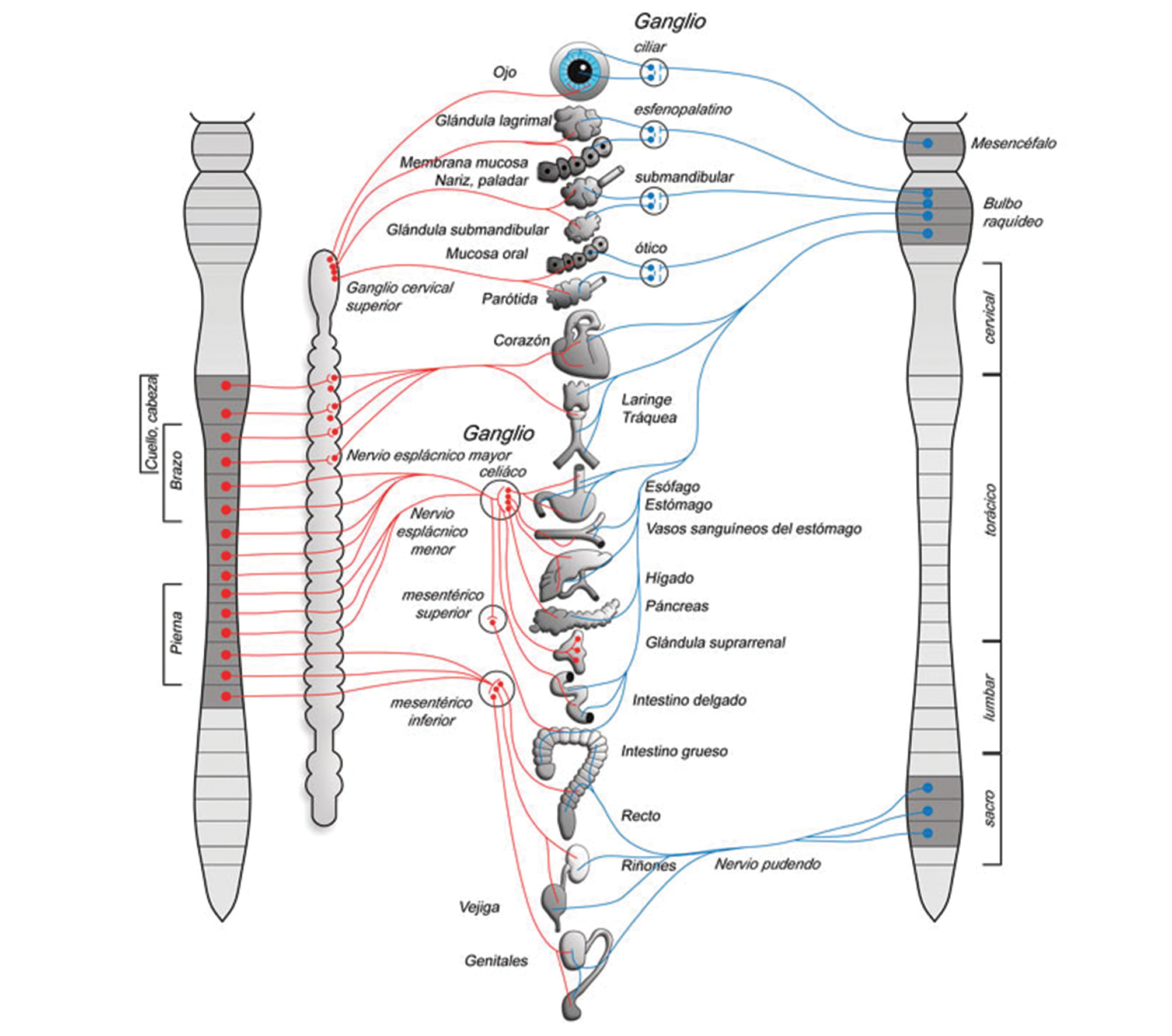
The peripheral part of the ANS consists essentially of the sympathetic and parasympathetic nervous systems. The sympathetic nervous system (red) originates from the thoracic medulla and the upper three segments of the lumbar medulla and is therefore also called the thoracolumbar system. The parasympathetic nervous system (blue) originates in the brainstem and sacral medulla and is therefore also called the craniosacral system.
CONTRADICTORY ORGANIC AND TISSUE REACTIONS CONTROLLED BY THE SYMPATHIC AND PARASYMPATHIC SYSTEMS
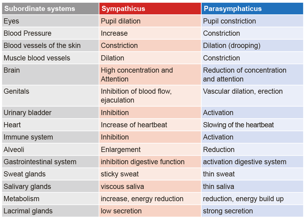
For example, if there is a long-term stress situation, the following values and laboratory parameters would be meaningfully increased, as the body is in a “fight and flight” response, i.e. a fight for survival:
- Increased blood pressure
- Accelerated heart rate
- Dilated pupils
- Increased energy production (ATP)
- Increased blood sugar level
- Activation of blood clotting
- Difficulty falling asleep and staying asleep
- Increased production of stress hormones
What are the possible false diagnoses and therapies without HRV analysis?
Physiological basics
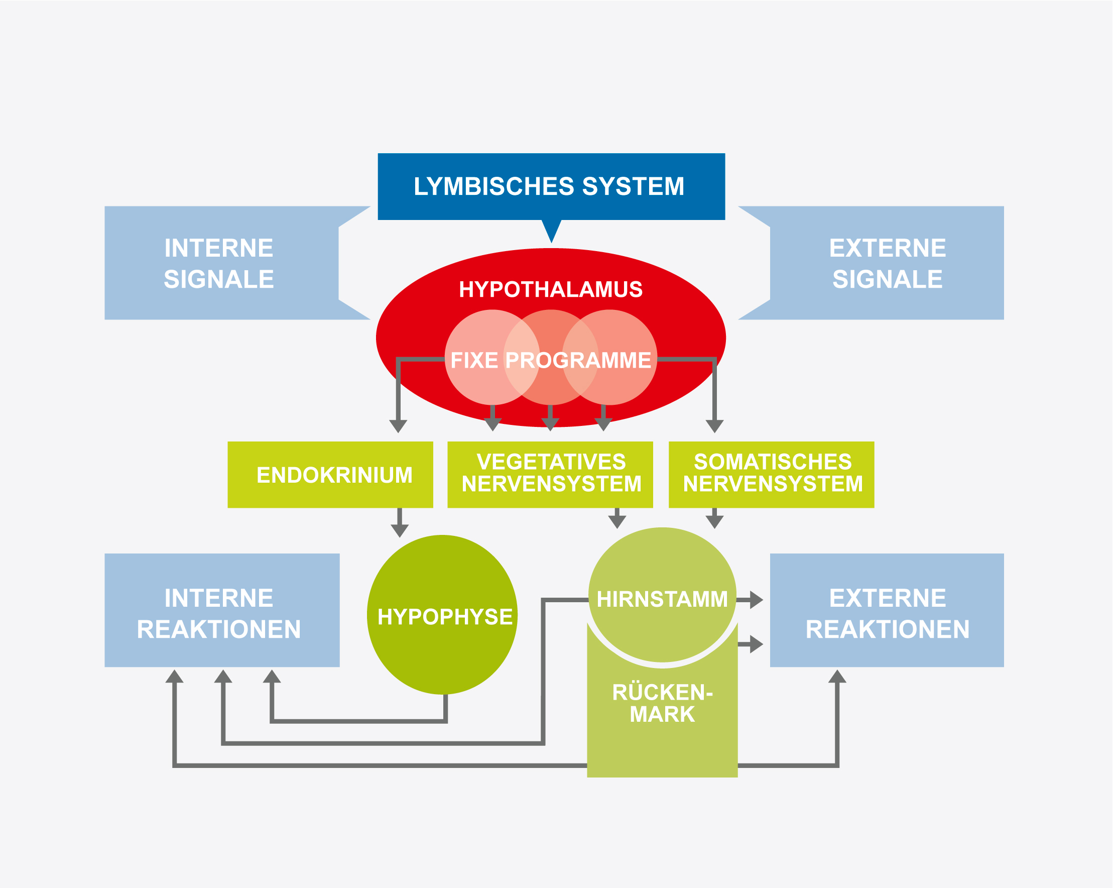
The autonomic nervous system (ANS), along with the central nervous system (CNS), is the most important neural control unit of the organism. Its main function is to adapt the internal milieu of the body to external and internal stresses (stimuli) and to maintain a constant function of the organism. The peripheral CNS is involved in a complex system that, in addition to connections to the brainstem, is also connected to the hypothalamus and other structures located in the CNS.
The scientist v. Hering prophesied in 1925:
“The wise use of the vegetative system will one day constitute the the main part of the medical art.”
The Limbic System
The limbic system, which is also called the mammalian brain, originated in the earliest stages of mammalian development. It controls and regulates the sensations important to the social aspects of mammals e.g. fear, love, desire, learning, concern for offspring and play instinct. The limbic system was first described by Paul Broca in 1878. The limbic system is not so much an organ in itself, but a functional unit responsible for processing emotion, drive and learning.
Control variables of HRV
The sinus node is located on the inner side of the posterior wall of the right atrium. The excitation originating from there is transmitted via the musculature to the atrioventricular node (AV node), and from there to the ventricles. The sinus node is the fastest and therefore superior pacemaker of the heart with an intrinsic natural frequency of 80-120 beats per minute.
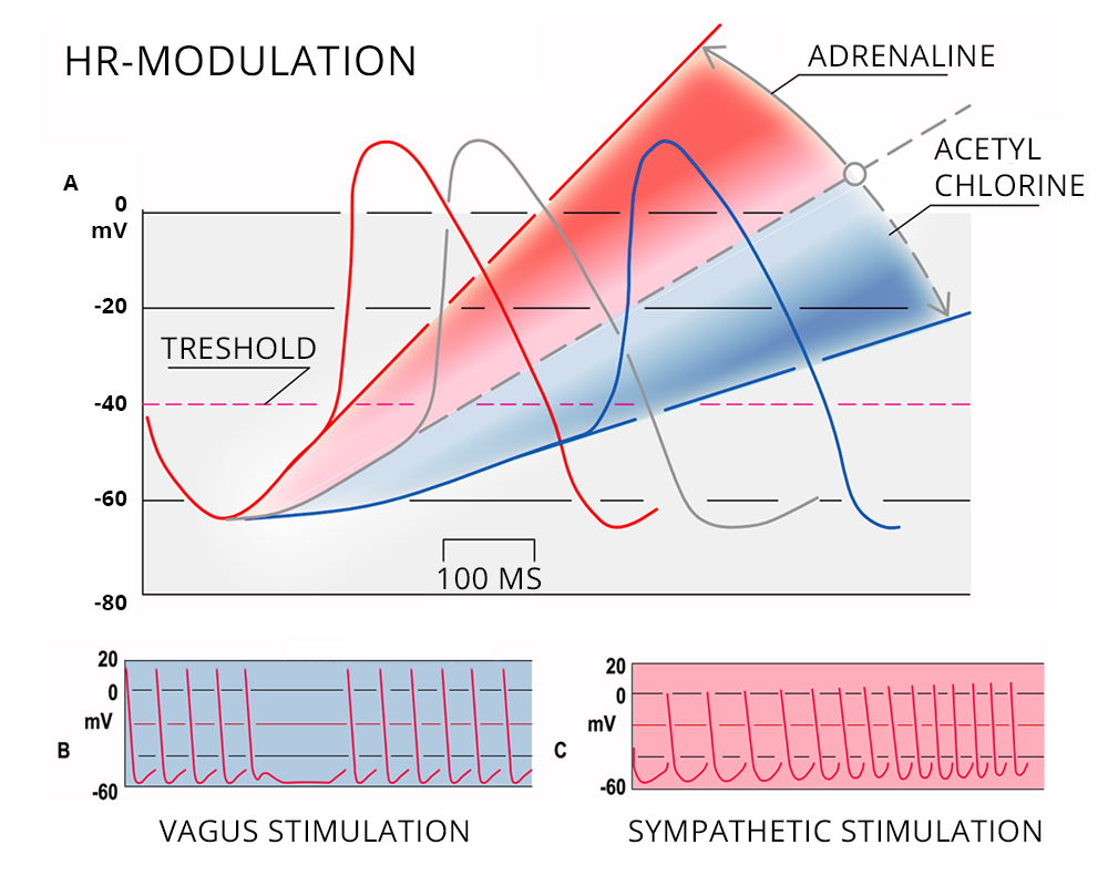
Heart rate modulation
The heart muscle is innervated by both sympathetic and parasympathetic parts. The sympathetic nervous system has a frequency-increasing (positive chronotropic) effect and the parasympathetic nervous system a frequency-decreasing (negative chronotropic) effect. If the heart rate is lower than the intrinsic excitation frequency of the sinus node (80-120), the parasympathetic nervous system is dominant and if this is higher, the sympathetic nervous system.
Baroreflex integration
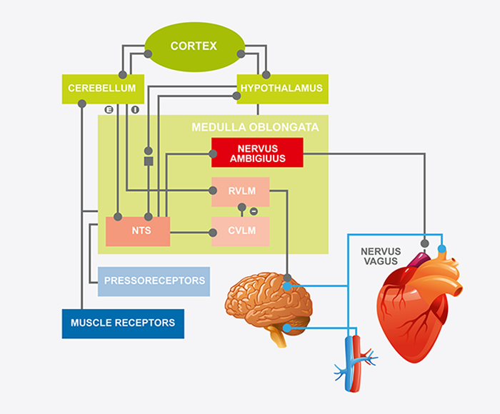
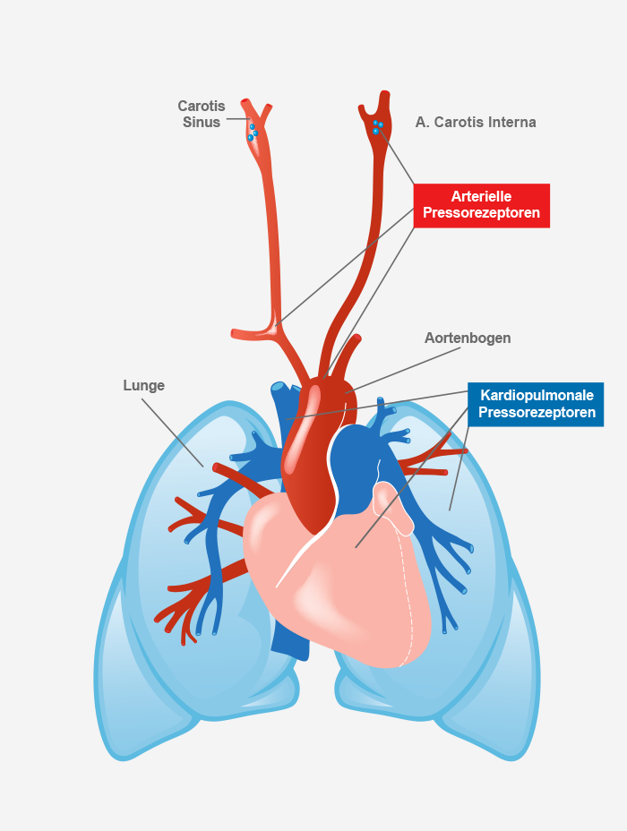
The signals from the baroreceptors reach the nucleus tractus solitarii (NTS) in the brainstem. From there, the signals reach the ambiguous nucleus and the rostroventrolateral and caudoventrolateral medulla oblongata, from which excitatory and inhibitory impulses are sent to the heart and vessels to control arterial blood pressure.
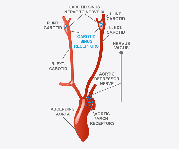
Baroreflex / Baroreceptors
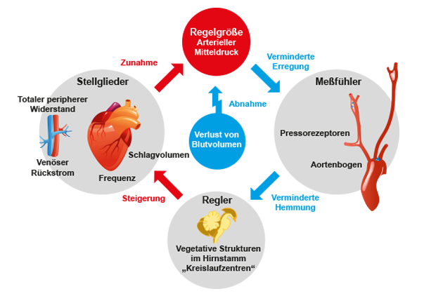
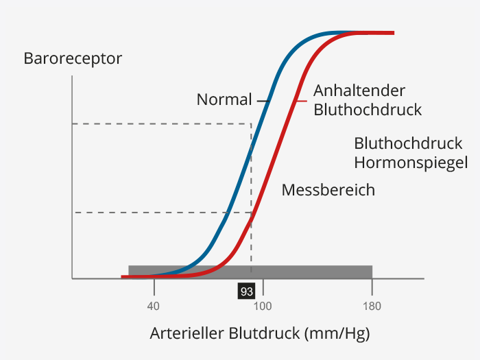
Baroreflex activity and respiratory sinus arrhythmia (RSA) are the central mechanisms influencing HRV. Baroreflex control serves to permanently maintain an adequate (mean) arterial blood pressure to supply all organ systems. The receptors located in the aortic arch and carotissinus have a very high sensitivity and react to minimal changes in pressure. During physical exertion or sport, there is a shift in the sensitivity threshold to meet the higher oxygen demand. An altered threshold due to permanent sympathetic stimulation plays a significant role in the development of hypertension.
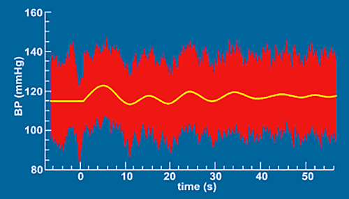
Respiratory sinus arrhythmia – RSA
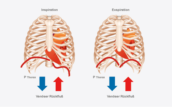
The dependence of heart rate on respiration is called respiratory sinus arrhythmia (RSA):
- During inspiration (inhalation), an increase in heart rate
- During expiration, a decrease in heart rate.
RSA is mediated primarily by the alternating activity of the vagus nerve.
Influences on respiration-dependent heart rate variability:
- Pulmonary, vascular, and cardinal stretch receptors. respiratory centers in the brainstem
- Different baroreflex sensitivity in the respective phases of the respiratory cycle.
Due to inspiratory vagal inhibition, fluctuations in heart rate occur at the same frequency as respiration.
Inspiratory inhibition is primarily caused by the influence of the medullary respiratory on the medullary cardiovascular center.
In addition, peripheral reflexes due to hemodynamic changes and thoracic stretch receptors are responsible.
RSA – a resonance phenomenon
The resonance phenomenon describes the superposition of, in this case, biological oscillations. The physiological respiratory sinus arrhythmia describes a frequency range of approx. 0.3Hz. By a clocked respiration with a frequency of 6 breaths/min. this frequency shifts into the range of approx. 0.1Hz, in which e.g. the basic frequency of the baroreflex integration is located. This achieves a change in HRV that can be directly displayed in the analysis. Fig. left Clearly visible in the rhythmogram: left HRV with “normal” breathing up to the middle of the rhythmogram, then HRV with clock breathing In the longer term, this breathing modulation leads to a strengthening of baroreflex control and improved activity of the parasympathetic nervous system.
Resipiratory sinus arrhythmia (RSA).
Resipiratory sinus arrhythmiaas a resonance phenomenon.
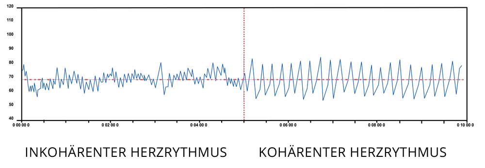
The rhythm of the heart can be spontaneously influenced by resipiratory pacing!
Spectral analysis – frequency-based parameters
The pure RR intervals can be decomposed into their frequency components by the FFT (Fast Fourier Transform). Frequency components in the ranges VLF, LF and HF result from the signal. The total power is given in TP (Total Power).
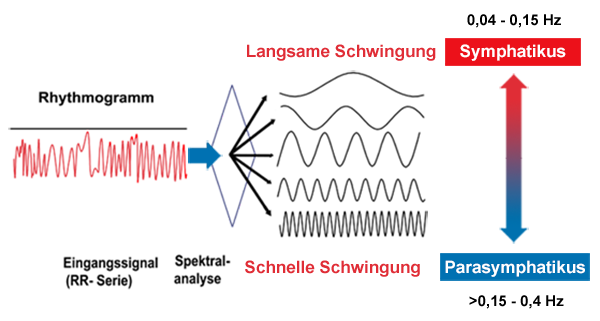
VLF – Very Low Frequency frequency range: 0.00 – 0.04Hz
LF – Low Frequency Frequency range: 0.04 – 0.15Hz
HF – High Frequency frequency range: 0.15 – 0.4Hz
LF/HF Ratio
From our point of view and that of renowned scientists and sports scientists, the frequency-based HRV parameters (spectral analysis) are not well suited for everyday use, as they are very susceptible and parameters such as the VLF range cannot be adequately represented in a short-term measurement. Therefore, we restrict ourselves to the time domain parameters that are valid for a short-term measurement.
Non-linear HRV parameters
Alpha 1 or DFA 1: detrended fluctuation analysis. The Alpha 1 value measures not only purely the temporal changes in heart rate variability, but it also measures the quality of regulation. These nonlinear parameters and values play a greater role in therapeutic application and evaluation.
By examining the raw HRV data for random and repetitive ranges, it is possible to analyze how the individual regulatory systems work together. Optimally, the alpha 1 value would be 1.0, which indicates that 50% of the signals in the heart rate variability are random, indicating rapid response, and 50% of the signals are repetitive. This indicates basic stability of the control systems.
Anything above 1.0 indicates more stability and is more likely to imply compensation processes in the individual control systems. Anything less than 1.0 means a lot of randomness and, from a value of less than 0.8, does not indicate good cooperation between the control systems. This condition can also be called chaos in the system.
For example, an example of stability in the system is the measurement under cycled breathing. When a measurement is performed under a given breathing rate, a respiratory sinus arrhythmia (RSA) occurs, which can be seen very nicely in the rhythmogram. The signal looks very uniform, which means there is more stability in the biological control system. The regulatory systems work very closely coupled with each other, there is the so-called coherence. The alpha 1 value therefore inevitably increases under cycle breathing. This is therefore not to be evaluated negatively but shows that the systems can enter into coherence.
The VNS analysis (the big brother of the HRV analysis) especially for therapists, practices and clinics with patient database, diagnosis and therapy database as well as other functions can be found here: Commit GmbH

